
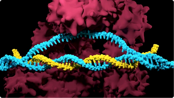
Disease modeling is a cornerstone of biomedical research, helping us understand how diseases develop, and how they can be treated. Over the past decade, advances in CRISPR gene editing have transformed disease modeling by enabling researchers to create more precise and biologically relevant models than ever before.
In this chapter, you will learn how model organisms are used in genetics research and why they remain essential tools for studying human disease. We will introduce the fundamentals of disease modeling, explore the major types of disease models and discuss the challenges and limitations associated with animal-based models. Finally, we will look at how CRISPR changed the field of model animal research, and how regulatory and industry shifts are shaping the future of disease modeling.
How do scientists understand what goes wrong in a disease, uncover new treatment strategies, and predict whether a therapy will work? Disease models provide the foundation for answering these questions.
Disease models are biological systems, including cells, tissues, and animals, that recreate key features of human diseases in the laboratory. They aim to capture the mechanisms behind disease development, and how diseases and therapeutics affect the rest of an organism. They try to capture a complete picture - the entire organism, and how drugs and diseases affect them - in a smaller, controlled manner.
There are several reasons why modeling diseases are important:
However, disease models are not perfect. Cell-based models cannot capture the interconnected complexity of organisms, while animal-based models are similar, but not perfect representations of human biology. Our immune system, reproduction, brain and nervous systems are only represented by animal models at a limited capacity.
Animal models are valuable tools for studying human disease, but many diseases are driven by genetic changes that are unique to humans. By introducing human genes or disease-associated mutations into an animal genome, researchers can create transgenic animals that more accurately mimic human disease biology and provide a more representative system for studying disease mechanisms and potential treatments.
The first attempts at creating transgenic animals were recorded in the early 1980s. Mice, a common model organism for genetics, were the first to have their genome modified. Early methods involved injecting a DNA fragment with the desired insert sequence into mouse embryos. This resulted in incorporation of the insert into random sites in the mouse genome, albeit at low efficiency (1–3). Although this achievement was a major advance in the field of genetics back then, the method had severe limitations such as high toxicity due to the off-target DNA inserts.
Over time, newer techniques, such as site-specific recombinases, ZFNs, and TALENS, improved the precision of editing specific genomic targets in animals. However, some issues persisted and these methods were inefficient and laborious, thereby limiting their scope of application - until CRISPR arrived.
Introduction of the CRISPR-Cas9 technology in 2012 was a game-changer in the field of genetic engineering. As CRISPR enabled targeted genome editing in a simple, efficient, and economical manner, the process of creating transgenic animals became simpler in the span of just a few years.
Moreover, commercial availability of synthetic single guide RNA (sgRNA) drastically reduced the experimental time and improved the editing efficiency when compared to in vitro transcribed (IVT) guide RNAs. For instance, Synthego’s synthetic sgRNA shows high editing efficiencies and high rates of germline transmission, key factors for successfully generating transgenic animals.
Development of CRISPR-based genome engineering technology enabled editing genes with such ease that standardized protocols for creating transgenic mice in the lab are now available for guiding researchers. The list of CRISPR-modified transgenic animals now extends far beyond mice to many more species. We will discuss some examples below.
Mice are among the most widely used animal models in biomedical research because they share many biological similarities with humans, including conserved genes, physiological pathways, and disease mechanisms. While not perfect replicas of human biology, their genetic and physiological similarities have made them invaluable tools for understanding our biology and diseases.
First studies into generating genetically engineered mice date back to 1974 with Rudolph Jaenisch and Beatrice Mintz, inserting the SV40 viral DNA into the germ line cells using microinjection. Throughout the years, genetically modified or selectively bred mice steadily became one of the most helpful models for the study of biology and human diseases. With the advent of CRISPR, it has become relatively simple to genetically modify mice and study mutant models.
The arrival of CRISPR genome editing dramatically accelerated the creation of genetically modified mouse models. Unlike earlier approaches that often required complex and time-consuming targeting strategies, CRISPR enables researchers to easily and rapidly edit mice genomes, introduce targeted mutations, delete genes, or insert new genetic sequences with greater speed and flexibility. With CRISPR, scientists can now generate mouse models that closely mimic human disease-associated mutations in a fraction of the time previously required.
Today, CRISPR-engineered mice continue to play a central role in biomedical research, from uncovering disease mechanisms to evaluating new gene therapies. Mice models continue to be pivotal in the fields of cancer biology, immunology, and neurodegenerative diseases. Their combination of genetic similarity to humans, well-established research tools, and compatibility with precise genome editing makes them one of the most important model systems for translating discoveries from the laboratory toward clinical applications.
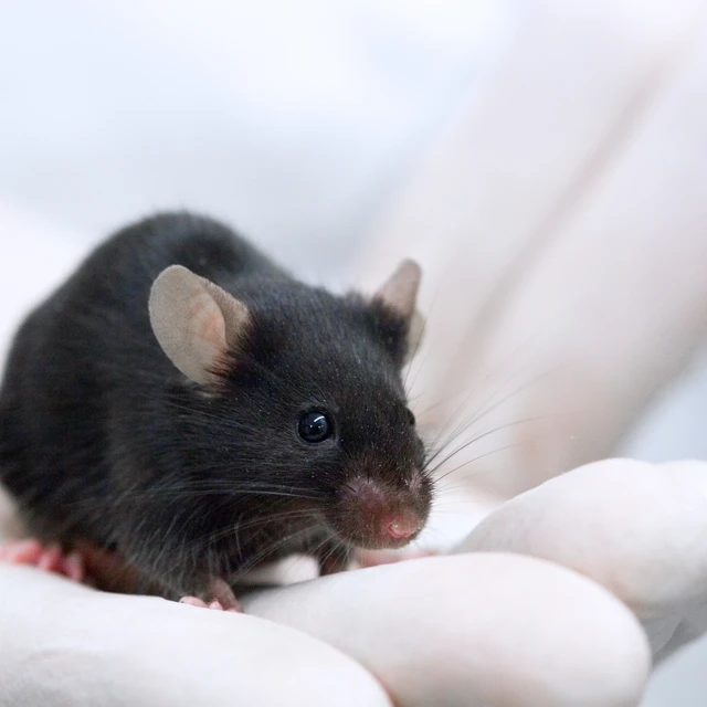
This CRISPR mouse embryo microinjection protocol, provided by researchers at the University of Rochester Mouse Genome Editing Resource, was designed to allow highly efficient introduction of synthetic single guide RNA into mouse embryos.
Since ancient times, the budding yeast Saccharomyces cerevisiae has been an essential part of our lives. Like bacteria, yeast is a simple, single-celled organism that is inexpensive and easy to grow. Unlike bacteria, however, yeast is a eukaryote, allowing you to study many cellular processes and metabolic pathways that are shared with human cells. One of the earliest genetic screening methods was the yeast-two-hybrid system, which helped determine if two individual proteins interacted with one another. With modern techniques, yeast models remain as one of our most valuable model organisms to study genetic pathways and biological processesthat cannot be modelled in simpler prokaryotes, such as DNA repair, cell division, vesicle trafficking, and metabolic pathways.
The development of CRISPR genome editing has further expanded the capabilities of yeast as a research model. Because yeast naturally has efficient mechanisms for repairing DNA through homologous recombination, researchers can use CRISPR to insert genes of interest to the yeast genome with high accuracy. By combining CRISPR with yeast’s rapid growth and simple genetics, scientists can quickly generate libraries of mutations and screen thousands of genetic changes to understand how specific genes influence cellular function. The benefits are also mutual - researchers have used yeast models to quickly screen CRISPR-Cas gRNAs to improve our understanding of gRNA design. From identifying gene functions to uncovering disease-associated pathways, CRISPR-engineered yeast models provide researchers with a fast, scalable, and cost-effective way to explore biology before moving discoveries into more complex models.
Zebrafish (Danio rerio) have become one of the most widely used vertebrate models for studying human biology and disease. Over 82% of genes associated with human diseases are found in Zebrafish (Danio rerio), making this small tropical freshwater fish a highly popular model for studying human diseases.
One of the key reasons behind this is their unique approach to reproduction and development. Imagine being able to watch an organ form, first beats of a heart, or see firsthand how a genetic mutation alter development. Zebrafish lay transparent, fertilized eggs, which makes it easy for researchers to inject gene editing agents directly into the eggs to modify developmental genes, and then study the results visually. The ability to observe development real-time make zebrafish a great model for studying bone, cardiovascular, and developmental diseases.
CRISPR has transformed this natural advantage into an even more powerful research tool. Because zebrafish embryos are easily accessible at the earliest stages of development, researchers can introduce CRISPR components directly into fertilized eggs and rapidly generate animals carrying specific genetic changes. Combined with advanced imaging techniques and high-throughput screening approaches, CRISPR zebrafish models are helping scientists uncover new disease mechanisms, study the effects of hazardous chemicals in development, and identify potential drug targets before advancing discoveries into more complex models.
Don't let its size fool you. Drosophila melanogaster, or the fruit fly, has become one of the most important model organisms in biology. Its scientific career began out of necessity in 1904, when Thomas Hunt Morgan was on the lookout for a cheaper and more convenient model than mice to study genetics. What started as a practical solution became a groundbreaking discovery platform, with Morgan’s experiments in fruit flies helping establish the chromosomal theory of inheritance and earning him the Nobel Prize in Physiology or Medicine in 1933.
Fruit flies have some unique genetic advantages that make them especially powerful research tools. Unlike many other organisms, male flies do not undergo meiotic recombination, which helps researchers maintain specific genetic combinations across generations. Researchers can also use their recessive lethal “balancer chromosomes” to maintain heterozygote populations.
The simplicity and speed of the fruit fly lifecycle make it an ideal system for large-scale genetic experiments. A single generation can be completed in just a few weeks, allowing researchers to rapidly study how genetic changes affect development, behavior, metabolism, and disease. Over the past century, these advantages allowed Drosophila models to contribute to discoveries in areas ranging from cancer biology and neurodegeneration to aging and immunity.
CRISPR has added a new level of precision to this already powerful genetic toolkit. By allowing researchers to directly modify specific genes, CRISPR enables the creation of fruit fly models carrying disease-associated mutations or engineered genetic changes with unprecedented speed and accuracy. Combined with the extensive genetic resources already available for Drosophila, CRISPR makes it possible to rapidly test gene function, investigate biological pathways, and uncover mechanisms underlying human disease.
At just about 1 mm in length, Caenorhabditis elegans may seem like an unlikely candidate for studying human biology. However, this tiny roundworm has become one of the most influential model organisms in genetics, developmental biology, and neuroscience. Since its introduction as a research model by Sydney Brenner in the 1960s, C. elegans has helped scientists uncover fundamental principles of gene regulation, cell development, aging, and programmed cell death.
One of the reasons C. elegans is such a powerful research tool is its remarkable simplicity and genetic accessibility. The worm has a fully mapped cell lineage, meaning researchers can trace the fate of individual cells from embryo to adult. Its transparent body also allows scientists to observe cellular processes in living animals without invasive procedures. Additionally, its short lifecycle, large number of offspring, and well-characterized genome make it possible to rapidly study how genetic changes affect an entire organism.
The marriage of CRISPR and C. elegans transformed the already convenient organism to a powerhouse of genetics research. Researchers can now engineer precise changes that mimic human genetic variants, allowing them to investigate how individual mutations influence development, cellular pathways, and disease-related processes. Combined with the worm’s simple biology and powerful genetic toolkit, CRISPR-engineered C. elegans continues to provide rapid insights into fundamental biology and human disease mechanisms.
Whether you are editing mice, zebrafish, insects, or emerging model organisms, successful genome engineering starts with the same foundation: carefully designed guide RNAs and reliable CRISPR reagents. From creating knockout models to introducing precise genetic changes, choosing the right CRISPR tools can help streamline your research and improve editing outcomes.
Explore our resources on generating CRISPR animal models, including guides and application notes covering gene knockouts, CRISPR delivery strategies, and successful genome editing approaches in animal systems. Got more questions? Our team of experts are here to help with all of your CRISPR needs.
Recombinase Polymerase Amplification (RPA) coupled with CRISPR-based detection offers a rapid, highly sensitive, and isothermal molecular diagnostic framework, bypassing the need for complex thermal cyclers. Operating at a constant 37°C...
Advancing a CRISPR program becomes more complex at every stage.
Cell-free protein expression (CFPE) systems enable rapid prototyping of protein constructs by expressing genes directly from engineered DNA templates in vitro. While linear templates allow fast iteration, circular DNA templates...
 Cart
Cart
 Account
 Your Orders
 Sign Out
 Sign In