
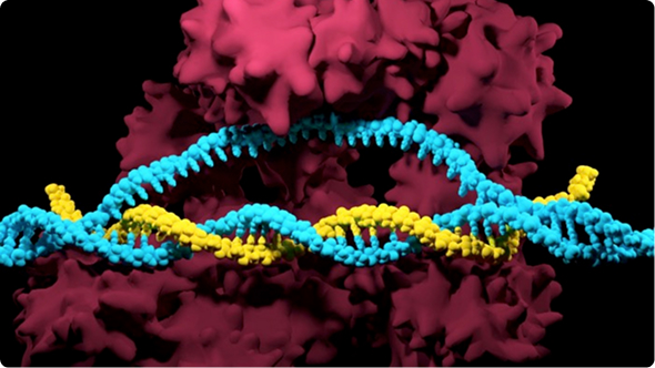
With the guide RNAs (gRNAs) and Cas proteins at hand, the last barriers ahead of genomic edits are the cell and nuclear membranes! In this chapter, we will cover how you can deliver your CRISPR system to your cells, types of CRISPR systems and how they affect delivery, and the advantages and disadvantages of various CRISPR delivery methods.
Once you've designed your guide RNA, the next step is delivering the CRISPR components into your target cells. For genome editing to occur, both the guide RNA and Cas nuclease must reach the cytoplasm or nucleus, depending on the delivery strategy. Introducing nucleic acids or CRISPR reagents into eukaryotic cells is known as transfection, and researchers have developed a wide range of methods to accomplish it.
The best transfection method depends on your experimental system and objectives. As you compare different approaches, there are five key factors to consider:
Let's take a deeper dive at how these factors may affect your decision in picking a transfection method for your experiments.
Whether you choose transient or stable transfection depends on how long you need your CRISPR components to remain active in your cells.
If you only need the Cas nuclease and guide RNA to be present temporarily, transient transfection is typically the preferred approach. Here, the CRISPR components are delivered into the cell without permanently integrating the DNA that encodes them into the genome. As a result, genome editing occurs only for a limited period before the CRISPR components are naturally degraded or diluted as cells divide. This shorter editing window helps reduce the risk of off-target effects, which is critical in developing clinical CRISPR therapeutics.
If your goal is to create a cell line that continuously expresses one or more CRISPR components, stable transfection may be a better choice. In this approach, the DNA encoding the Cas nuclease, guide RNA, or both is permanently integrated into the cellular genome, allowing these genes to be passed on to daughter cells during cell division. Depending on your experimental design, expression can be continuous (constitutive) or activated only in response to a specific stimulus, such as a small-molecule inducer (inducible). Stable transfection is most commonly achieved using viral vectors, although plasmid DNA can occasionally integrate into the genome at low frequency.
A popular strategy is to first generate a stable Cas9-expressing cell line. Once established, you can introduce different guide RNAs by transient transfection whenever you want to edit a new target, allowing you to reuse the same cell line across multiple experiments without repeatedly generating a stable line.
Because stable transfection relies on permanent genomic integration, it requires considerably more time and effort than transient approaches. Researchers typically include a selectable marker, such as an antibiotic resistance gene, enrich for successfully modified cells, and isolate individual clones for validation before beginning downstream experiments. For this reason, stable transfection is generally reserved for applications that require long-term or repeated CRISPR activity across multiple cell generations.
The format of your CRISPR components may affect your decision of which transfection method to use. Guide RNA and Cas9 can be delivered to cells as DNA, RNA, or as pre-formed ribonucleoprotein complexes (RNPs) formats.
Transfection efficiency often depends on cell type. Most common cell lines and immortalized cells are generally easy to transfect. Primary cells and stem cells, on the other hand, are more sensitive and often have lower viability after transfection.
Immortalized cell lines propagate indefinitely in laboratory cell culture conditions. They are either derived artificially by inducing mutations that prevent senescence, or naturally from cancers. Because immortalized cells divide frequently (during which the nuclear envelope is broken down), there are ample opportunities for CRISPR components to enter the nucleus. Thus, these cell lines are generally easy to transfect. One drawback of cell lines is that they can accrue genetic mutations over time, and these changes can be significant.
Because of this, cell lines may be aneuploid and are less representative of the natural biology of living tissues. They are also vulnerable to contamination. However, because they are cost-effective and easy to culture and manipulate, immortalized cells are commonly used in research, especially for early proof-of-concept experiments.
Examples: HEK293, HeLa, A549, Jurkat
Primary cells are derived directly from living tissue without introducing additional genetic changes. Primary cells are subdivided into two types: adherent (requiring attachment to a surface in culture) or suspension (not requiring attachment). The ability of primary cells to propagate is limited and therefore, they cannot be cultured for extended periods of time.
Because primary cells have limited numbers of cell divisions, there are fewer opportunities for CRISPR components to enter the nucleus. Thus, these cells are typically difficult to transfect. However, primary cells may undergo a process called transformation, either naturally or following stimulation, in which they gain mutations that make them immortalized. A significant benefit of primary cells is that they are more representative of the natural biology of living tissues than immortalized cells.
Examples: Adherent cells from organs (kidney, liver, epithelial cells, fibroblast), suspension cells from blood (T cell, lymphocyte)
Stem cells have natural properties that enable them to propagate in an undifferentiated state for an indefinite amount of time. When cultured in the lab, they can either be maintained as undifferentiated cells or stimulated to differentiate. Stem cells can either be pluripotent (able to differentiate into any cell type) or multipotent (able to differentiate into some cell types). The main types of stem cells are embryonic stem cells (ES cells), adult stem cells, and induced pluripotent stem cells (iPS cells).
Examples: iPS cell, ES cell, Hematopoietic stem cells (HSC)
Germline cells develop into gametes (sperm and eggs) and can be transfected before or after fertilization. In sexually-reproducing organisms, a sperm fertilizes an egg, producing a single-celled zygote. The zygote then divides and develops into a multicellular embryo. Gametes and embryos are generally difficult to transfect by most methods.
Examples: Mouse pronucleus (the nucleus of a fertilized egg), insect eggs, embryos
Your choice of a CRISPR transfection protocol may depend on the number of samples you need to process at a time. For large numbers of samples, a high-throughput method is preferable. These methods often involve the use of automated equipment or more advanced machinery. Alternatively, a low-throughput method may be adequate for smaller numbers of samples. Some sample types, such as mouse embryos, can only be efficiently transfected using low-throughput methods.
The transfection method you choose may also depend on the equipment and reagents you have available. Some delivery methods require specialized instruments, such as an electroporator or nucleofector, along with compatible buffers and consumables, while others only require standard laboratory equipment and transfection reagents.
When selecting a delivery method, consider whether your lab already has access to the required equipment or whether purchasing new instrumentation fits your budget and research plans. If you expect to perform CRISPR editing routinely, investing in specialized equipment may be worthwhile. For occasional experiments, a method that relies on more widely available reagents and instruments may be the more practical choice.
For most CRISPR applications, we need to deliver our CRISPR-Cas complexes to the nucleus of our cells to edit their genomes. This is achieved by the use of transfection methods - physical, chemical, or viral-mediated ways to deliver the CRISPR-Cas complex across cellular barriers. Each method has different advantages and disadvantages with respect to efficiency, throughput, equipment, skill, and cost.
|
Method
|
Principle
|
Best For
|
Advantages
|
Limitations
|
Compatible Cargo
|
CellCompatibility
|
Typical Applications
|
|
Electroporation
|
An electric pulse creates temporary pores in the cell membrane, allowing CRISPR cargo to enter.
|
Suspension cells and many primary cells
|
High delivery efficiency; supports DNA, RNA, and RNP delivery
|
Requires optimization; can reduce cell viability
|
DNA, mRNA, sgRNA, RNP
|
Broad range of cell types
|
Ex vivo cell engineering, pooled screens
|
|
Nucleofection
|
A specialized form of electroporation that delivers cargo directly to the nucleus using cell type-specific electrical programs.
|
Difficult-to-transfect primary cells, stem cells, immune cells
|
Excellent efficiency in difficult cell types; optimized protocols available
|
Requires specialized instruments and proprietary reagents; higher cost
|
DNA, mRNA, sgRNA, RNP
|
Broad range, including sensitive primary cells
|
CAR-T engineering, stem cell editing, primary cell editing
|
|
Microinjection
|
A microneedle directly injects CRISPR components into individual cells, oocytes, or embryos.
|
Embryo editing, transgenic animal generation
|
Precise delivery with minimal cargo loss; very high editing efficiency
|
Labor-intensive, low throughput, requires significant technical expertise
|
DNA, RNA, RNP
|
Individual cells, embryos, oocytes
|
Animal model generation, embryo editing
|
|
Lipofection
|
Lipid nanoparticles encapsulate CRISPR cargo and fuse with the cell membrane.
|
Easy-to-transfect adherent cell lines
|
Low cost, simple workflow, scalable, high throughput
|
Lower efficiency in primary cells; some cell types are resistant
|
DNA, mRNA, sgRNA, RNP
|
Mostly immortalized cell lines
|
Routine CRISPR knockouts, plasmid transfection
|
|
Viral Delivery
|
Viral vectors package CRISPR cargo and deliver it through natural infection mechanisms.
|
In vivo delivery and long-term expression
|
Efficient delivery in vitro and in vivo; enables stable or transient expression
|
Packaging size limits, biosafety considerations, longer experimental timeline
|
DNA, RNA (vector-dependent)
|
Broad range, depending on viral tropism
|
Gene therapy, in vivo genome editing, stable cell line generation
|
Physical transfection protocols create temporary holes in the plasma membrane through which gRNA/Cas9 can pass. Three popular physical methods to introduce CRISPR components into cells are electroporation, nucleofection, and microinjection. Electroporation and nucleofection use an electrical pulse to create pores in the plasma membrane, while microinjection uses a needle to force a hole through the membrane.
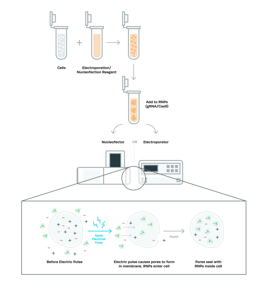
Electroporation involves suspending cells in a conductive solution and briefly applying high-voltage electrical pulses. The application of electricity induces the formation of temporary pores in the plasma membrane and the electrical potential across the membrane causes charged molecules (e.g., gRNA/Cas9) to enter the cytoplasm through the pores. Nucleofection is based on electroporation, but utilizes a special machine, called a Nucleofector, and has several distinctions from traditional electroporation (discussed below). Electroporation and nucleofection are effective for transfection of many cell types.
|
Advantages
|
Disadvantages
|
|
Fast and easy
|
Requires specialized equipment
|
|
High efficiency
|
Cell death may result from high voltage pulses and incomplete membrane repair
|
|
Large numbers of cells can be transfected in a short time (minutes)
|
Traditional electroporation collects the cells, gRNA/Cas9, and reagents in a pipette tip or a specialized electroporation cuvette. The electrical pulse is then applied to the components to facilitate the introduction of the CRISPR components across the plasma membrane, and sometimes also into the nucleus.
There are only a few buffers available that are compatible with electroporation, and each sample must be processed one at a time (low-throughput). A benefit of this device is that it is an open system and the user can manually set the parameters for each sample. This characteristic allows one to optimize specific parameters for each cell type. A popular electroporation device is ThermoFisher Scientific’s Neon Transfection System.
Nucleofection uses a Nucleofector (Lonza) machine designed to facilitate the transfer of gRNA/Cas9 directly to the nucleus. The procedure involves combining target cells, a buffer specific to each cell type, and gRNA/Cas9. These components are transferred to a cuvette or 16-well strip, which is then placed into a Nucleofector. An electric pulse with parameters pre-optimized for each cell type is then applied.
The specificity of the solution and electric pulse enables the gRNA/Cas9 to directly enter the nucleus. Because cell division is not required for the transfer into the nucleus, nucleofection is an effective method for non-dividing cells (e.g., neurons, blood cells). Multiple samples can be transfected at once, making it a higher throughput method than traditional electroporation. Nucleofection with RNPs is the preferred method to introduce the CRISPR components into most cell types in terms of editing efficiency.
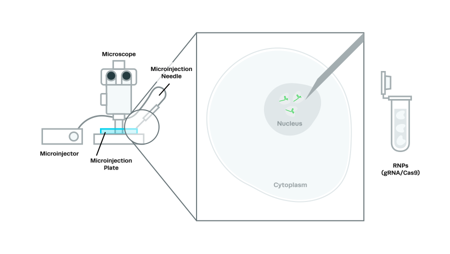
Microinjection involves carefully positioning a target cell under a microscope and delivering gRNA/Cas9 into the cytoplasm or nucleus through a glass micropipette. This method is highly technical and requires special equipment, including micromanipulators and a microinjector. It is also low-throughput, as only one cell can be transfected at a time.
However, this method can result in very high efficiency when performed by skilled individuals. Microinjection is mainly used for transfecting single cells, oocytes, zygotes, and embryos. It is a popular method for pronuclear injections (into the nucleus of fertilized eggs), which is used to generate transgenic mice and other animals.
|
Advantages
|
Disadvantages
|
|
High efficiency (near 100%)
|
Labor-intensive & requires skill
|
|
Dosage control
|
Low-throughput
|
|
Low cytotoxicity
|
Requires special equipment
|
Several chemically-based methods can be used to transport molecules into cells, including calcium phosphate, cationic polymers, and cationic amino acids. One of the most common methods to introduce CRISPR components into cells is lipofection.
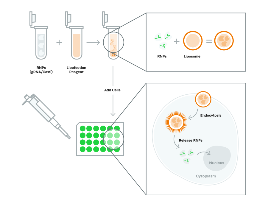
Lipofection (also called lipid-based transfection) uses cationic lipid reagents to deliver CRISPR components into cells. This method first involves constructing lipid-soluble structures, called liposomes, around the CRISPR components. The components are then transported into the cell through endocytosis, a process in which the plasma membrane surrounds the components on the exterior-side, and buds off inside the cell. The CRISPR components then escape the endosomal pathway and diffuse through the cytoplasm. Unlike nucleofection and microinjection, lipofection does not deliver CRISPR components to the nucleus.
Lipofection is an easy and economical method that usually has minimal toxicity. However, lipofection efficiency is usually not as high as the other methods This method is not recommended for primary cells or stem cells.
|
Advantages
|
Disadvantages
|
|
Economical & easy
|
Moderately efficient
|
|
Can be adapted to high-throughput systems
|
Limited cell types
|
|
Does not require specialized equipment
|
No delivered directly to the nucleus
|
Viral vectors can be used to transfer DNA or RNA into cells in a process called transduction. The process first involves packaging the gRNA/Cas9 sequences into viral particles and then introducing the particles into target cells. To make the viral particles, the plasmid containing the gRNA or Cas9 sequence and plasmids containing viral genes are introduced into a packaging cell line (e.g., 293 T cells). Once the viral particles are produced, they are harvested from the packaging cells and introduced into the target cells that one wants to transfect with gRNA or Cas9.
There are several types of viruses that can be used for transduction, including lentivirus, adenovirus, adeno-associated virus, and herpes viruses. For stable transduction, lentiviruses, a family of retroviruses that includes HIV, are often used because they integrate their genome into the genome of infected cells (Fig 4). Lentiviruses can penetrate the nuclear envelope, and thus are effective in non-dividing cells (i.e., it is not reliant on mitosis for nuclear entry). Lentiviruses are effective in a variety of cell lines, primary cells, and stem cells, and can be used in vivo. However, because they integrate randomly into the genome, they may disrupt vital genes or alter the regulation of gene expression.
Adeno-associated virus (AAV) is another type of virus commonly used for in vivo editing and often results in the sustained expression of exogenous genetic sequences in dividing and non-dividing cells. AAVs typically do not trigger an immune response and have a range of serotypes that enable targeting to different tissues. Another benefit of recombinant AAVs is that they do not integrate into the genome, and will not cause disruption of genes through insertional mutagenesis.
One drawback of AAVs is their limited packaging capacity of about 4.5 kb, so it may be a challenge to fit both gRNA and Cas9 sequences in a single vector. AAVs are also commonly used to transport donor DNA templates into cells for knock-in experiments.
Viral vectors are highly efficient at transfecting CRISPR components and compatible with many cell types. However, constructing viral particles is time-consuming and laborious. Depending on the type of virus used, extra safety measures may be required in the laboratory.
|
Advantages
|
Disadvantages
|
| High efficiency |
May require extra safety measures |
| Compatible with many cell types | Laborious & time-consuming |
| Can be used in vivo and in vitro |
Some types may have immunogenicity & cytotoxicity |
There isn’t a “one-size-fits-all” answer to what protocol everyone should use for their CRISPR experiments. The sample type and experimental goals will primarily determine the best protocol for each experiment, and in every case, the protocol selected will require optimizations to achieve the highest editing efficiencies.
Visit the resources section of our website to check out our complete list of CRISPR protocols and other guides to help you achieve the highest level of success in your genome engineering experiments.
Congratulations! You have introduced all the necessary CRISPR-Cas9 components into your cells! Now, you want to know if it worked. Validating your CRISPR editing is an extremely important step in your experiment, and there are many options available to assess CRISPR editing efficiency.
In the next chapter, we will look at the different methods you can use to analyze your cells to evaluate your CRISPR editing efficiency.
CRISPR has quickly become a standard laboratory tool for gene editing. As the adoption of CRISPR accelerates worldwide, up-to-date knowledge of the basics of CRISPR is essential for anyone in...
CRISPR has ignited a revolution. Although it’s a relatively recent discovery in the history of biotechnology, CRISPR has quickly become a standard laboratory tool and is transforming cell engineering-based research....
Confused about chemical modifications on CRISPR guides? Join us as we explore why chemical modifications are so important, the different types and their applications, and how Synthego’s modified guides can...
 Cart
Cart
 Account
 Your Orders
 Sign Out
 Sign In